
Figure 3. Monocytes and macrophages in the development of an atherosclerotic lesion.
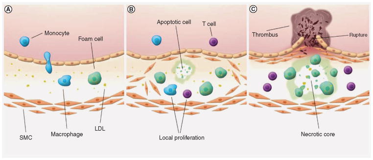
(A) The initial processes of atherosclerotic plaque initiation are unknown. However, early inflammation in the arterial wall results in the infiltration of monocytes that differentiate into activated macrophages. Macrophages will take up oxidized LDL and free lipids, transforming into lipid-rich foam cells. (B) A deregulation of the inflammatory process causes increased expression of adhesion molecules on endothelial cells above, resulting in enhanced influx of immune cells. At the same time, SMCs are thought to grow outward and establish themselves under the endothelial layer, producing extracellular matrix proteins that eventually form a fibrous cap. Local proliferation of macrophages and lymphocytes increases atherosclerotic plaque growth. (C) As more immune cells promote the inflammation, foam cells and SMCs go into apoptosis and form a necrotic core. Proteases produced by macrophages further destabilize the lesion, which is believed to increase the risk of plaque rupture. A ruptured lesion exposes its tissue factors to coagulation factors in the blood, resulting in the formation of a thrombus. LDL: Low-density lipoprotein; SMC: Smooth muscle cell.