
Fig. 1.
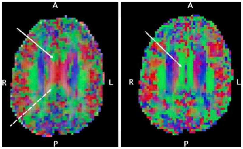
Axial-oblique (parallel to AC-PC line) DTI color maps of the brain at level of (left) and above (right) the upper lateral ventricles from a selected participant. Red denotes fibers in the left-right direction, green denotes fibers in the anterior-posterior direction, blue denotes fibers in the inferior-superior direction. In the left panel, the DTI voxel selection of the anterior site of the cingulum white matter tract (solid white arrow) and posterior site of the cingulum white matter tract (dashed white arrow) are shown. These are the relatively small anterior-posterior oriented tracts mesial and anterior/posterior respectively to the corpus callosum as it forms a prominent “X” at the level of the upper lateral ventricles. In the right panel, the DTI voxel selection of the right middle cingulum site (dot-dashed white arrow) on a supraventricular axial-oblique slice is shown, centered in the midcaudally situated anterior-posterior tract just mesial to the superior corona radiata.