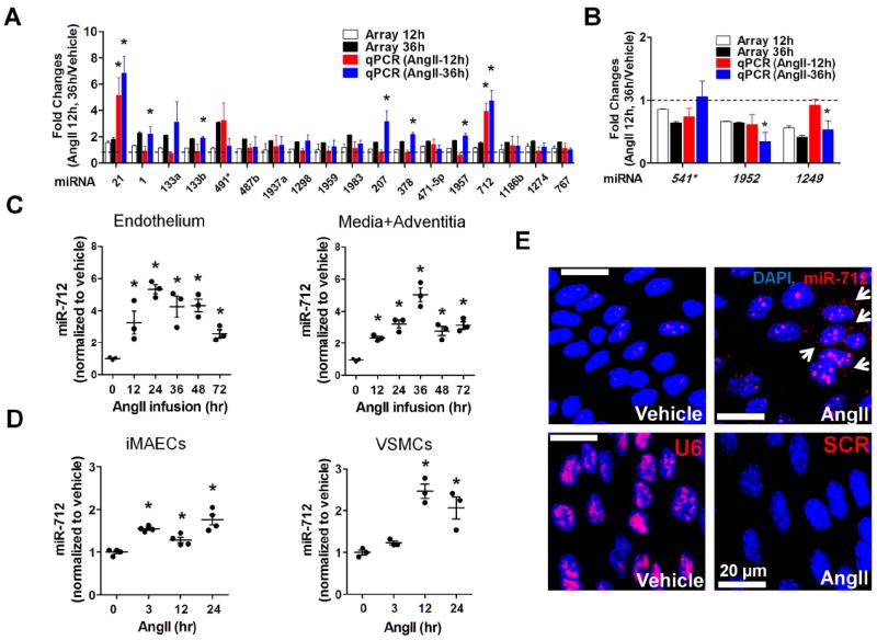
Figure 1. Identification and validation of AngII-sensitive miRNA, miR-712 in in vivo and in vitro.

(A, B) Endothelial-enriched total RNAs, obtained from the abdominal aorta (C57BL/6 mice) at 12 and 36hr post-AngII infusion (1 μg/kg/min), were analyzed by miRNA array and quantitative PCR (qPCR). Twenty one miRNAs (18 up-, 3 down-regulated miRNAs at 36 hr post-AngII implantation compared to the vehicle control) were selected based on fold-change (18 up-regulated miRNAs : >1.5 fold and 3 down-regulated miRNAs : <0.65 fold) and statistical significance (p<0.05) in the miRNA array study. These miRNAs were further validated by qPCR and their fold-change over the vehicle control are shown along with the microarray results (compared to vehicle control; mean±S.E.; *p<0.05). Data were analyzed by Student’s t-test. (C) Expression of miR-712 was determined by qPCR using endothelial-enriched RNA from AngII-infused abdominal aorta and leftover RNA (media/adventitia) obtained from AngII-infused C57BL/6 mouse (0-72hr). (D) miR-712 expression was tested in immortalized mouse aortic endothelial cells (iMAECs) and vascular smooth muscle cells (VSMC) in vitro (0-24hr). (C, D : Data were analyzed using ANOVA followed by Tukey’s post hoc test, mean±S.E. *p<0.05; n=4). (E) Abdominal aortas of C57BL/6 mice obtained at 2-days post AngII-infusion were subjected to fluorescence in situ hybridization using digoxigenin-labeled miR-712 probe (red) and confocal microscopy, (n=4). Blue: DAPI nuclear stain; Arrows indicate cytosolic miR-712.
