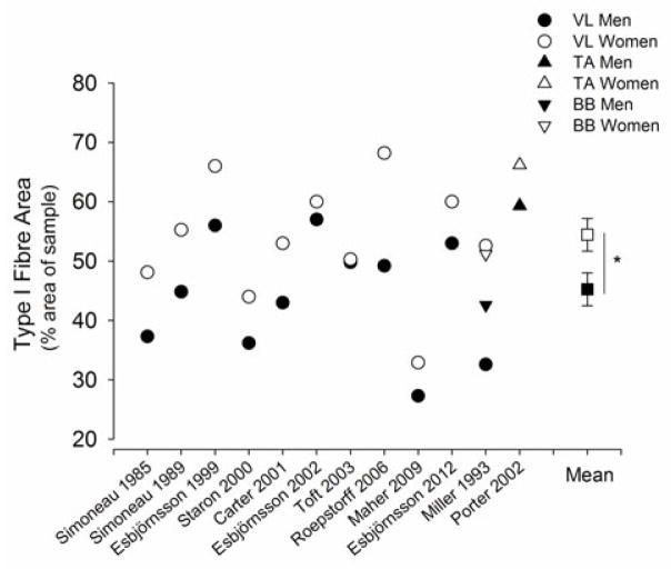
Figure 5.
Type I fibre area (%, proportional area) of skeletal muscle histochemically analysed for myosin ATPase activity from muscle biopsy samples of vastus lateralis (VL), tibialis anterior (TA) and biceps brachii (BB) in young men (closed symbols) and women (open symbols) that were sampled in the same study. Shown are the mean proportional areas of the men and women in each of the 12 studies (Simoneau et al., 1985, Simoneau and Bouchard, 1989, Miller et al., 1993, Esbjornsson-Liljedahl et al., 1999, Staron et al., 2000, Carter et al., 2001b, Esbjornsson-Liljedahl et al., 2002, Porter et al., 2002, Toft et al., 2003, Roepstorff et al., 2006, Maher et al., 2009, Esbjornsson et al., 2012). The mean (± SEM) per cent area of type I fibres of all the muscles from the 12 studies is plotted on the right side. Women had greater type I fibre area (%) than men (P<0.05).

