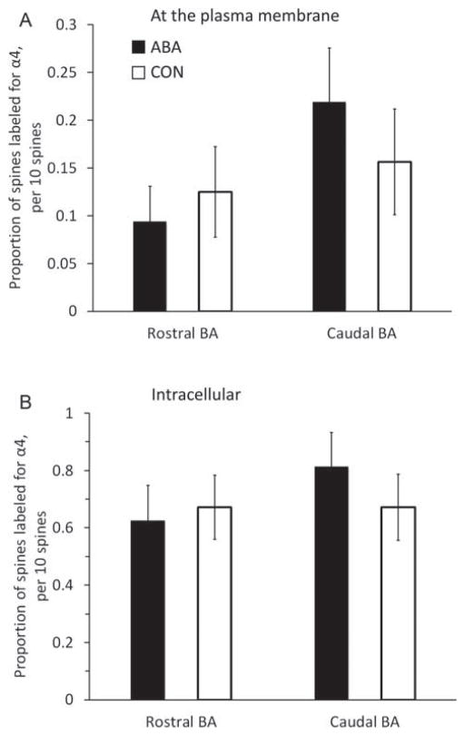
Fig. 3.
Comparisons of immunoreactivity for the α4 subunit within dendritic spines of the rostral and amygdala of females following 4 days of treatment. We compared the levels of α4 immunoreactivity (-ir) in the rostral and amygdala from brains of control animals (CON; n = 4), and activity-based anorexia animals (ABA; n =4). The proportion of α4-ir spine profiles encountered was measured. To this end, for every group of 10 spines that was randomly encountered, the number of spine profiles immunolabeled at the plasma membrane was assessed. This assessment of the proportion of spine profiles labeled was repeated 16 times for a single source of tissue, to obtain a mean value of 16 assessments, representing the analysis of 160 spine profiles. Any single spine profile was categorized as labeled at the membrane, so long as it contained one or more SIG particles at the plasma membrane. The 16 assessments from each animal were pooled group-wise, resulting in 64 values in each group for the rostral and caudal amygdala (four animals per group). In a parallel manner, we also measured the proportion of spine profiles immunolabeled intracellularly for the α4 subunit. As described for the membranous label analysis, an assessment of 160 spines per animal yielded 64 values in each group for the rostral and caudal amygdala (four animals per group). There was no difference between the groups by Mann–Whitney U test, either in the membrane counts (Panel A) or intra-cellular counts (Panel B). Error bars denote standard errors of the mean.

