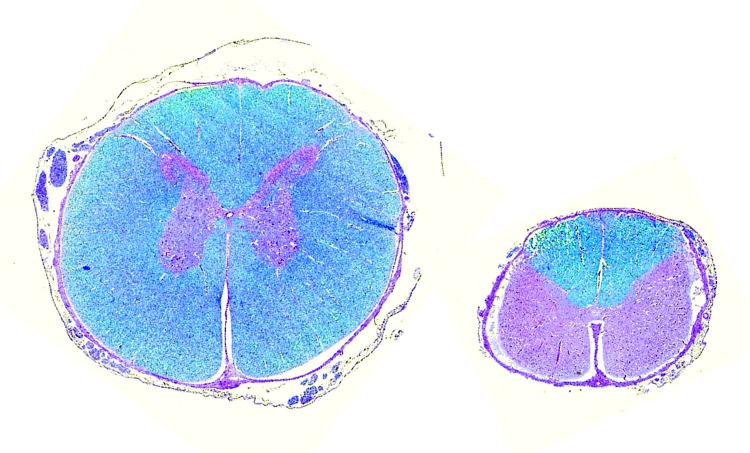
Figure 2.
Micromyelia. Age- and site-matched spinal cord transversal histologic section at the level of C4. Left, control calf; right, Schmallenberg virus (SBV)–infected calf. Note atrophy/hypoplasia and prominent deficiency of stainable myelin in ventral and lateral tracts of SBV-infected calf. Luxol fast blue staining.

