
Figure 5.
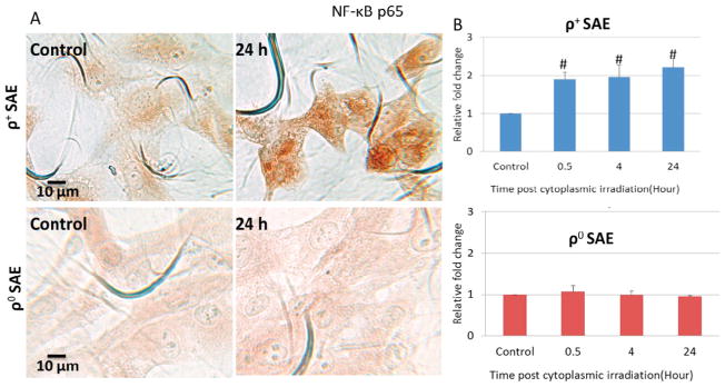
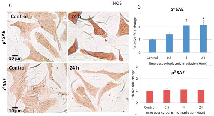
Effects of NF-κB in the regulation of genomic instability induced by cytoplasmic irradiation in ρ+ SAE cells. A. Representative image of NF-κB p65 immunocytochemistry staining of ρ+ and ρ0 SAE cells. B. Quantification of NF-κB p65 by mean gray value of ρ+ and ρ0 SAE cells. Levels of NF-κB p65 were significantly increased post cytoplasmic irradiation with ten α particles in ρ+ SAE cells but not in ρ0 SAE cells. C. Representative image of iNOS immunocytochemistry staining of ρ+ and ρ0 SAE cells. D. iNOS expression were significantly increased induced by cytoplasmic irradiation in ρ+ SAE cells, however, ρ0 SAE cells showed no response.
#, P<0.01, *, P<0.05 versus the control group. Bars indicate ± SD. Results were repeated in three other experiments. In each experiment, 100 cells were scored.