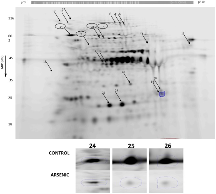
Figure 6. 2DE analysis of proteins from T. asahii labeled with IAF at protein thiols.
Prior to 2DE, protein thiols were labeled with IAF. Gels were scanned for IAF-associated fluorescence and images were analysed by Progenesis Samespot analysis software. Images were normalized prior to applying any statistics. Spots showing significant (p<0.05) decrease in IAF-associated fluorescence were excised for identification and are indicated by arrows (see Table 2). Inset: Zoom boxes are shown for spots.

