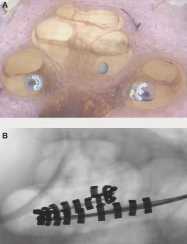
Figure 6.

Human temporal bone showing (A) the perimodiolar position of the Contour Advance inserted using AOS; (B) a cross-sectional, high-resolution radiograph of the human temporal bone. (Images by permission of the Cooperative Research Centre for Cochlear Implant and Hearing Aid Innovation, Melbourne, Australia.)
