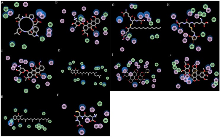
Figure 4. Two-dimensional representations of the 10 reference compounds, showing H-bond and hydrophobic interactions within substrate binding sites of ABCC6 open conformation.
(A) BQ-123, (B) Doxorubicin, (C) Daunorubicin, (D) Vitamin K1, (E) vitamin K2, (F) S-2(2, 4-dinitrophenyl) glutathione, (G) Leukotriene C4, (H) NEM-GS, (I) Teniposide, and (J) Etoposide are shown respectively. Residues involved in Van der Waals interaction are represented by green disks, residues involved in polar interaction or hydrogen bonding are represented by pink disks and solvent accessible surfaces of the interacting residues are represented by a blue halo around the residues.

