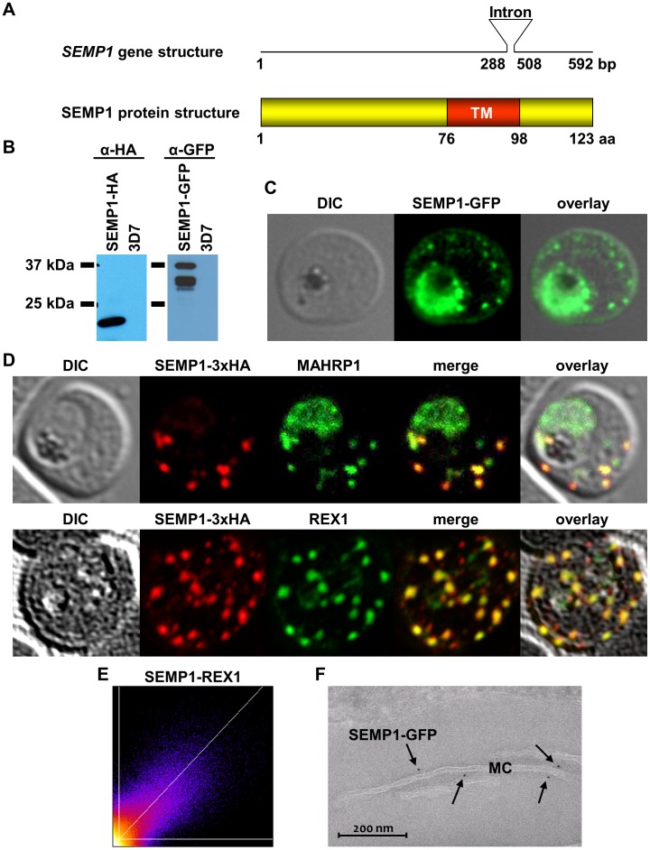
Figure 1. SEMP1 expression and localization.
A: schematic representation of the semp1 gene (top) and SEMP1 protein structure (bottom). TM, transmembrane protein. B: Live cell imaging of 3D7 parasites expressing SEMP1 with C-terminal tagged GFP. C: Immunofluorescence assays of MeOH-fixed RBCs infected with 3D7 expressing SEMP1 with a C-terminal 3xHA tag, co-labelled with rat α-HA and either rabbit α-MAHRP1 or rabbit α-REX1 antibodies. D: Scatter plot of co-localization of SEMP1 and REX1 in SEMP1-3xHA parasites. E: Electron microscopy (EM) of RBCs infected with 3D7 expressing SEMP1 with a C-terminal GFP tag labelled with rabbit α-GFP antibodies and decorated with 5 nm gold conjugated Protein A.

