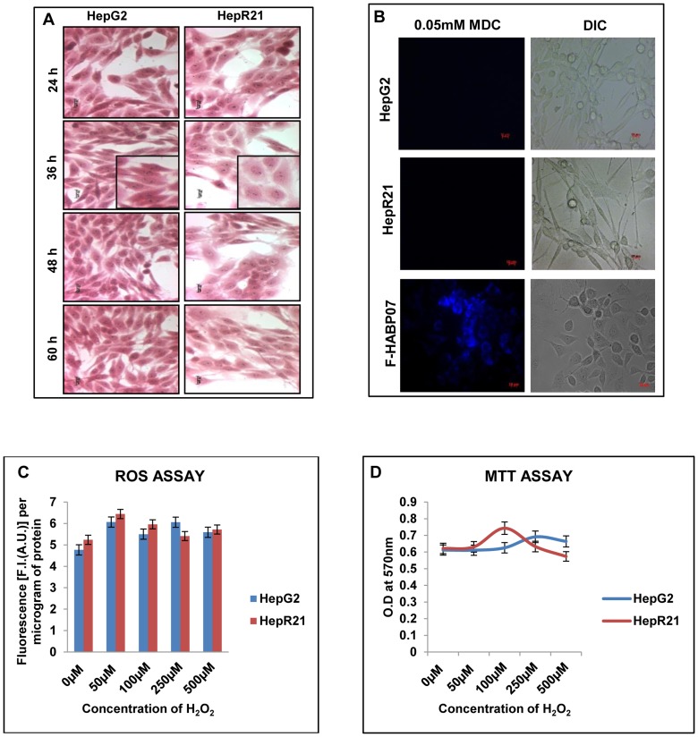
Figure 1. Morphology of HepG2 and HepR21 remains unaffected on progression of growth with both cell lines showing redox insensitivity.
[A] Haematoxylin-Eosin Staining — Morphological analysis by H-E staining of HepG2 and HepR21 cells illustrated that HABP1 transformed HepG2 i.e. HepR21 although morphologically varied than its normal counterpart having a higher cell volume, with a more flattened spread out appearance showed no change in morphology as a function of time. Increased generation of vacuolated HepR21 cells upon progression of time point was not observed. A magnified view of both the cells for 36 h of growth has been shown. [B] MDC staining shows absence of autophagic vacuolation in both HepG2 and HepR21 cells — HepG2 and HepR21 cells were grown for different periods starting from 36 to 84 h and subjected to MDC (0.05 mM) staining as per the protocol mentioned in Methods. Subsequent fluorescence microscopy indicated no positive staining for autophagic vacuoles for any point of growth between 36 h to 84 h. MDC staining of F-HABP07 cells was taken as positive control. Scale bar represents 10μ. [C] HepG2 and HepR21 cells are both insensitive to external redox stimuli — ROS assay performed on HepG2 and HepR21 cells after prior treatment of cells with varying concentration of H2O2 (0 µM, 50 µM, 100 µM, 250 µM and 500 µM) for one hour indicated both HepG2 and HepR21 cells to be redox insensitive even on exposure to 500 µM of H2O2. [D] H2O2 treatment has no effect on survivability of HepG2 and HepR21 as evident from MTT Assay — HepG2 and HepR21 cells were grown in complete media till 48 h and then treated with the abovementioned concentrations of H2O2 for 1 h. Viability assay performed thereafter revealed that even 500 µM of H2O2 has no effect on the survivability of both the cell lines. The media was not changed at any point.

