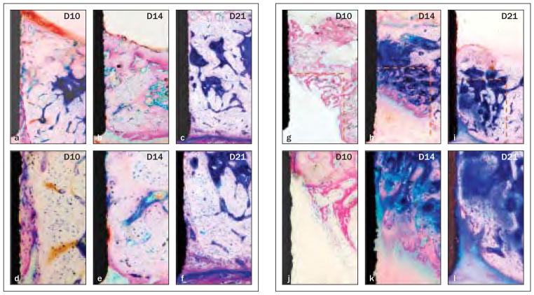
Fig 4. Bone apposition from the base and lateral walls of osteotomy over time (methylene blue and acid fuchsin; original magnification ×40 in the top row, ×200 in the bottom row).
Figs 4a to 4f Defects in the OA group. At day 10 (left-hand column), a 0- to 50-μm gap (dashed line in the first column) on the interface was filled by new bone. Significant maturation of the interfacial tissue was evident (middle column) on day 14 and (right-hand column) on day 21.
Figs 4g to 4i Defects in the OS group. (Left-hand column) A woven trabecular bone structure grew from the border of the defect at day 10. Significant bone maturation and apposition were noted (middle column) on day 14 and (right-hand column) day 21.

