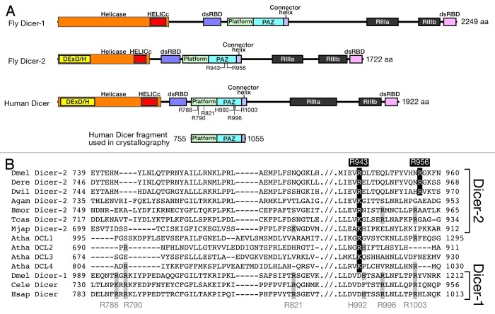
Figure 1. Dicer domain architecture. (A) Domain structures of fly and human Dicers. DExD/H, DExD/DExH box helicase domain; HELICc, helicase conserved C-terminal domain; dsRBD, dsRNA-binding domain; PAZ, PAZ domain; RIIIa and RIIIb, Ribonuclease III domain. The fragment of human Dicer containing the platform domain, the PAZ domain, and the connector helix, whose crystal structure was determined in complex with dsRNA (Fig. 5)19 is also shown. (B) Alignment of the platform and PAZ domain sequences from Dicers from various arthropods, Arabidopsis, C. elegans, and humans. Dmel, Drosophila melanogaster; Dere, Drosophila erecta; Dwil, Drosophila willistoni; Agam, Anopheles gambiae; Bmor, Bombyx mori; Tcas, Tribolium castaneum; Mjap, Marsupenaeus japonicus; Atha, Arabidopsis thaliana; Cele, Caenorhabditis elegans; Hsap, Homo sapiens. In this review, we number human Dicer amino acid residues based on its 1922-aa full-length sequence, whereas in the Park et al. and Tian et al. papers6,19 and the PDB files therein, the amino acid residues are numbered based on 1,912 aa sequence, which is lacking the first 10 aa compared with the 1922 aa sequence.

An official website of the United States government
Here's how you know
Official websites use .gov
A
.gov website belongs to an official
government organization in the United States.
Secure .gov websites use HTTPS
A lock (
) or https:// means you've safely
connected to the .gov website. Share sensitive
information only on official, secure websites.
