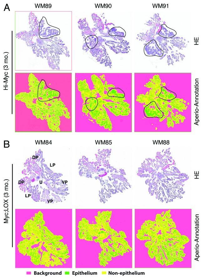
Figure 2. Inhibition of PIN and tumor development in 3-mo-old Myc;LOX transgenic mice. (A) WM prostates from 3 representative 3-mo-old Hi-Myc animals (total n = 15). Shown are HE (top) and the corresponding Aperio ScanScope (below) images. The areas demarcated by black lines indicate the PIN and early adenocarcinoma lesions. The WM numbers are indicated on top. (B) WM prostates from 3 representative 3-mo-old Myc;LOX dTg animals (total n = 7). Shown are HE (top) and the corresponding Aperio ScanScope (below) images. The orientation of the WM is illustrated on the HE image of WM84. U, urethra; DP, dorsal prostate; LP, lateral prostate; VP, ventral prostate. The virtual slides shown below were generated with Aperio ScanScope with trainable GENIE morphometric software that permits morphometric quantification of scanned images. The color coding was indicated below. Note that the histological presentations of WM91 and WM88 at higher magnifications are shown in Figure 1C.

An official website of the United States government
Here's how you know
Official websites use .gov
A
.gov website belongs to an official
government organization in the United States.
Secure .gov websites use HTTPS
A lock (
) or https:// means you've safely
connected to the .gov website. Share sensitive
information only on official, secure websites.
