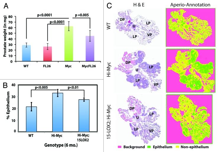
Figure 4. 15-LOX2 suppresses Hi-Myc adenocarcinoma in 6-mo-old dTg animals. (A) Prostate weight comparisons of WT and Tg animals. DLP and VP lobes of mice (n ≥ 7) from respective genotypes were dissected out, and collective weights of the lobes measured. (B) Quantification of epithelial tumor areas. Shown are the mean epithelial area (%) quantified from WM prostate sections in WT (n = 5), Hi-Myc (n = 7) and Hi-Myc;15-LOX2 (n = 5) mice using the ScanScope images. (C) Representative H&E images of samples used for quantifications in (B) and the virtual slides generated with Aperio ScanScope with trainable GENIE morphometric software that permits morphometric quantification of scanned images. For HE sections, the 4 prostatic lobes and urethra (U) were indicated. For the virtual slides (right), the color coding was indicated below.

An official website of the United States government
Here's how you know
Official websites use .gov
A
.gov website belongs to an official
government organization in the United States.
Secure .gov websites use HTTPS
A lock (
) or https:// means you've safely
connected to the .gov website. Share sensitive
information only on official, secure websites.
