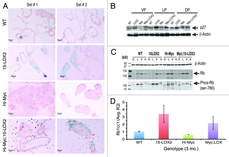
Figure 6. Cell senescence in Myc;LOX prostates. (A) Representative SA-βgal staining images in 3-mo-old prostates showing senescence in the Myc;LOX prostates. (B) Western blot analysis showing p27 expression in various prostate lobes of 3-mo mice among different genotypes. β-actin was used as the loading control. (C) Altered Rb1 and Phos-Rb in Myc;LOX prostates. Prostatic lobes were microdissected out from the animals of the indicated genotypes (3 mo) and used in western blotting analysis of the indicated molecules. (D) Increased Rb1cc1 mRNA expression in 15-LOX2 single or double transgenic prostates. Presented are the mean ± SD data from 2 sets of mice.

An official website of the United States government
Here's how you know
Official websites use .gov
A
.gov website belongs to an official
government organization in the United States.
Secure .gov websites use HTTPS
A lock (
) or https:// means you've safely
connected to the .gov website. Share sensitive
information only on official, secure websites.
