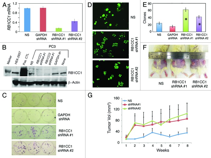
Figure 9. RB1CC1 knockdown promotes PC3 cell clonal and tumor growth. (A) Confirmation of RB1CC1 knockdown by qRT-PCR. PC3 cells were transduced with non-silencing (NS), GAPDH, or 2 RB1CC1 shRNA lentiviral vectors, and cells were then selected with puromycin to generate stable cell lines, from which total RNA was isolated for qRT-PCR analysis of RB1CC1 mRNA. (B) Confirmation of RB1CC1 knockdown by western blot analysis using an antibody from Sigma (# SAB4200135). Both PC3 and HEK-293T cells express endogenous RB1CC1 protein (~200 kD). The positive control (Pos. CTL) lane were HEK293T cells transfected with an RB1CC1-overexpressing plasmid. (C–E). RB1CC1 knockdown promotes PC3 cell clonal growth. The 4 types of stably selected PC3 cells were plated at clonal density (100 cells/well) in 6-well plates and images were captured at the end of 3 d (D) or 2 wk (C; giemsa stain). Shown in (E) is quantification of clones at the end of one week (mean ± S.D; n = 3). *P < 0.001 (compared with NS or GAPDH conditions). (F and G) RB1CC1 knockdown promotes PC3 xenograft tumor growth in NOD/SCID mice. The 3 types of stably selected PC3 cells as shown were subcutaneously injected (500 000 cells/injection) into dorsal flanks of 6-wk-old NOD/SCID mice (n = 10 injections/cell type), and the volume measurements were taken on a weekly basis (G). Shown in (F) are representative tumor images of each cell type.

An official website of the United States government
Here's how you know
Official websites use .gov
A
.gov website belongs to an official
government organization in the United States.
Secure .gov websites use HTTPS
A lock (
) or https:// means you've safely
connected to the .gov website. Share sensitive
information only on official, secure websites.
