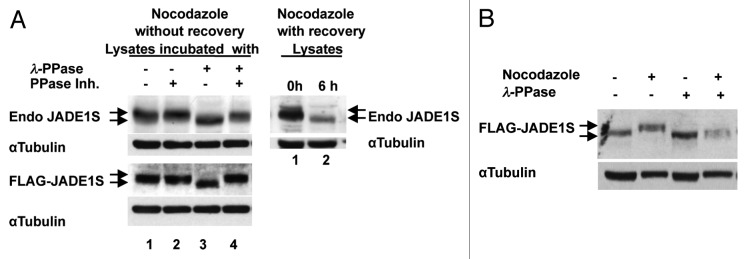
Figure 6. Nocodazole-dependent JADE1S gel band shift collapses after treatment with λ-phosphatase. (A) Left panel: lysates from synchronized HeLa cells enriched with high molecular weight specie of endogenous (upper panel) or overexpressed (lower panel) JADE1S (lane 1) were incubated with phosphatase (lane 3), phosphatase inhibitors (lane 2) or both (lane 4). Right panel: lysates from cells synchronized with nocodazole (lane 1) and after cell cycle release (lane 2) as in Figure 4. Endogenous and overexpressed JADE1S proteins were analyzed by western blot with JADE1S or FLAG antibodies. (B) JADE1S protein from cells treated with nocodazole but not vehicle collapses after treatment with phosphatase. JADE1S and HBO1 cDNA were co-transfected into H1299 cells, lysates incubated with phosphatase as in (A). Note that, protein amounts analyzed were adjusted for better bands resolution and do not represent the effect of nocodazole on JADE1S protein enrichment in soluble fraction.
