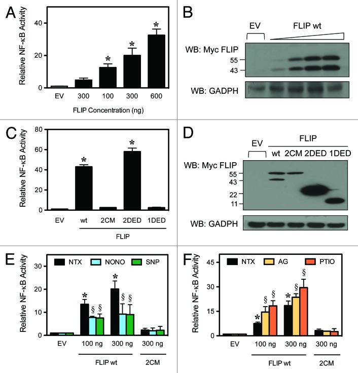
Figure 2. FLIP-mediated NF-κB activation. HEK-293 cells were transiently transfected with an NF-κB-luciferase and Renilla-luciferase reporter and expression constructs for the FLIP domains, as indicated. Luciferase activity was determined by the dual-luciferase reporter assay. (A) Cells were transiently transfected with increasing amounts of FLIP wt and an NF-κB luciferase reporter plasmid. NF-κB activity was determined by luminescence according to “Materials and Methods”. (B) Total cell lysates were also analyzed by western blot using an anti-myc antibody to verify the increasing expression levels of FLIP. (C) Cells were transiently transfected with empty vector (EV), FLIP wt, FLIP 2CM, FLIP 2DED, or FLIP 1DED and analyzed for NF-κB activity as noted in (A). (D) Total cell lysates were also analyzed by western blotting as above to verify expression of all constructs. (E) Cells were transiently transfected with EV, FLIP wt or FLIP 2CM. Cells treated with media alone, 400 μM NONOate, or 500 μM SNP for 12 h to supplement nitric oxide and analyzed for NF-κB activity as noted in (A). (F) Cells were transiently transfected with EV, FLIP wt, or FLIP 2CM, and cells were treated with media alone, 300 μM AG or 300 μM PTIO for 12 h to inhibit NO production. Lysates were analyzed for NF-κB activity as noted in (A). Data are mean ± SD (n = 3). *P < 0.05 vs. non-treated (NTX) EV-transfected cells. §P < 0.05 vs. non-treated (NTX) control.

An official website of the United States government
Here's how you know
Official websites use .gov
A
.gov website belongs to an official
government organization in the United States.
Secure .gov websites use HTTPS
A lock (
) or https:// means you've safely
connected to the .gov website. Share sensitive
information only on official, secure websites.
