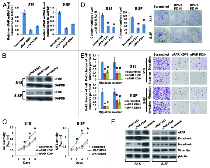
Figure 2. uPAR suppression affects the EMT and inhibits growth, colony formation, migration, and invasion in NPC cells. The highly metastatic S18 and 5-8F cells were transduced with lentiviruses expressing scrambled shRNA or shRNAs targeting uPAR at different loci (KD#1 and KD#4). uPAR mRNA levels (normalized to GAPDH) determined by quantitative real-time PCR (A) and uPAR protein levels determined by immunoblotting (B) in uPAR knockdown cells were both significantly reduced. (C) uPAR suppression impedes the growth of S18 and 5-8F cells as determined by MTS assays. *P < 0.01 for uPAR KD#1 and KD#4 compared with the scrambled controls. (D) uPAR suppression reduces the number of colonies formed with S18 and 5-8F cells. *P < 0.0001 relative to the scrambled controls. (E) uPAR suppression remarkably attenuates S18 and 5-8F cell migration and invasion as evaluated by the Transwell assays. *P < 0.001, #P < 0.05 compared with the scrambled controls. Photomicrographs are 100× (right panel). (F) Immunoblotting of EMT markers reveals elevated E-cadherin expression as well as decreased N-cadherin and vimentin expression upon uPAR suppression in NPC cells (vimentin expression in 5-8F cells was not affected by uPAR suppression). The data are presented as the mean ± SD of triplicate replicates.

An official website of the United States government
Here's how you know
Official websites use .gov
A
.gov website belongs to an official
government organization in the United States.
Secure .gov websites use HTTPS
A lock (
) or https:// means you've safely
connected to the .gov website. Share sensitive
information only on official, secure websites.
