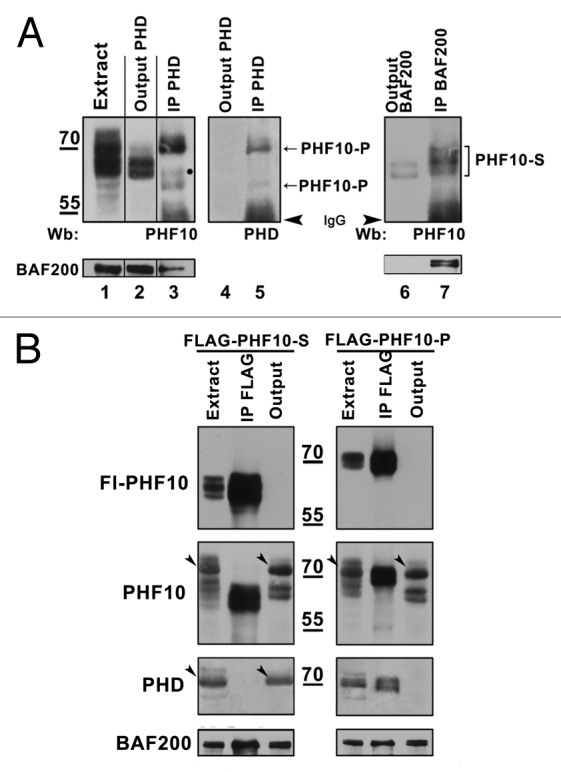
Figure 4. PHF10 isoforms containing PHD fingers or PDSM motif are associated with distinct PBAF complexes. (A) PBAF complexes containing distinct PHF10 isoforms are present in the HEK293 cell extract. The PHF10-P associated PBAF complexes were depleted from the extract with anti-PHD antibodies (lanes 1–5), while the complexes that contained PHF10-S remained in the output material and were further precipitated with anti-BAF200 antibodies (lanes 6–7). The western blot was developed with antibodies against PHF10 (lanes 1–3, 6, 7) or anti-PHD (lanes 4, 5). A significant amount of BAF200 co-precipitated with each of the PHF10 isoforms (lower panel), suggesting that PBAF complexes with PHF10-P or PHF10-S were both well represented in the cells. (B) Recombinant FLAG-tagged PHF10-S (left panel) or PHF10-P (right panel) in stably transfected HEK293 cells do not co-precipitate with endogenous PHF10-P and PHF10-Sl isoforms. The western blot was developed with anti-FLAG, anti-PHF10, and anti-PHD antibodies. Each recombinant isoform (panel FLAG-PHF10) was completely depleted from the extract, while the endogenous isoform with a different C-terminal domain remained in the output material (panels PHF10, PHD). Endogenous isoforms PHF10-P (left panel) and PHF10-Sl (right panel) are indicated by arrowheads.
