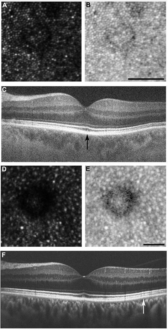
Figure 6.
Phenotypes 2 and 3: Decreased waveguiding and disrupted photoreceptor mosaic. Drusen were present in four subjects in which the overlying photoreceptor mosaic was undisturbed, consistent with previous observations.11 Interestingly, the photoreceptors on the edge of these drusen had diminished reflectivity. This is most likely due to the Stiles-Crawford effect; where the three dimensional druse alters the trajectory of the reflected light. (A) AOSLO image from subject JC_0803, displaying decreased waveguiding phenotype. (B) Logarithmic view of AOSLO image. Scale bar = 50 µm. (C) Corresponding OCT, with region imaged with AOSLO marked with vertical black arrow. In three subjects, drusen were present and the overlying photoreceptor mosaic was disturbed. By viewing on a logarithmic scale, it is evident that the photoreceptors are no longer discernable on the edge of drusen. These photoreceptor changes were not detected on SD-OCT. (D) AOSLO image from subject JC_1018, displaying disrupted photoreceptor mosaic. (E) Logarithmic view of AOSLO image. Scale bar = 50µm. (F) Corresponding SD-OCT, with region imaged with AOSLO marked with vertical white arrow.

