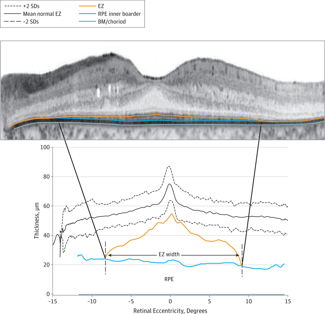
Figure 1.
Spectral-Domain Optical Coherence Tomography Scan of the Horizontal Midline in a Patient With X-Linked Retinitis Pigmentosa
Segmentation lines for the inner segment ellipsoid zone (EZ; inner/outer segment border) line band, the outer segment–retinal pigment epithelium (OS-RPE) junction, and the Bruch membrane (BM)–choroid junction are superimposed on the scan. The OS thickness relative to the normal mean EZ (2 SDs) for 23 healthy subjects is shown below. Vertical dashed lines indicate the retinal eccentricities at which OS thickness drops to 0 (ie, EZ becomes indistinguishable from the RPE).

