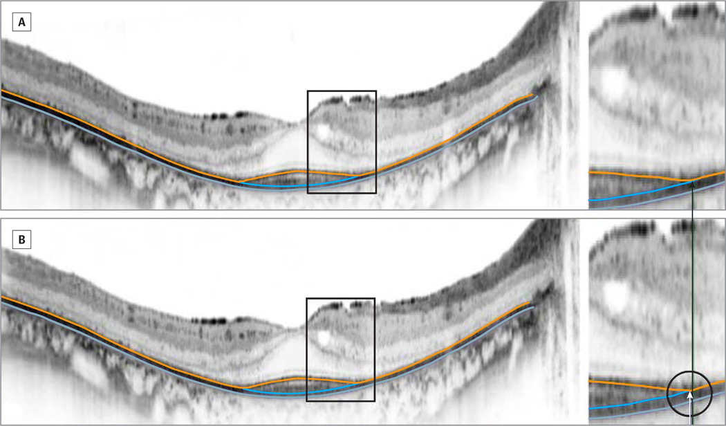
Figure 2.
Spectral-Domain Optical Coherence Tomography Scans of the Horizontal Midline From a Patient With Autosomal Recessive Retinitis Pigmentosa Undergoing Scanning Twice on the Same Day
A and B, Magnified views of the transition zone are shown to the right. The first and second scans differed in inner segment ellipsoid zone (inner/outer segment border) width by 0.3° (86 µm).

