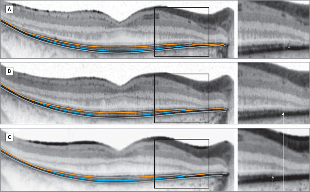
Figure 5.
Representative Scans From a Patient With X-Linked Retinitis Pigmentosa Across 3 Annual Visits
At the right is shown the inset at higher magnification. Arrows indicate the nasal edge of the inner segment ellipsoid zone (EZ; inner/outer segment border) each year. We detected a progressive shift in the remaining EZ band toward the center during the 3 annual visits.

