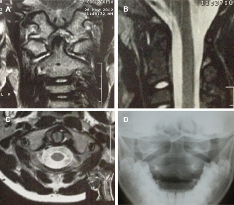
Fig. 5.

Magnetic resonance of the cervical spine, T2 sequence, after spontaneous reduction of the lesion. Observe the joint effusion on the anterior and posterior facets of the dens on the axial image. Intact transverse ligament of the axis (A). Joint effusion anterior to the dens on the sagittal image (B). Note the joint effusion in the atlantoaxial joint in the coronal image (C). Anteroposterior radiograph taken after 6 months demonstrates a reduced atlantoaxial joint (D).
