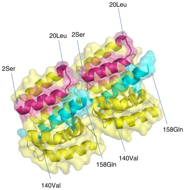
Fig. 5.
Structure of dimeric N-terminal domain (2–158 AA) of the M1 protein. Crystal Structure of the N-terminal domain (2–158 AA) of M1 protein is from influenza A virus (PDB = 3MD2). The structure was constructed using Pymol and the surface is shown as transparent. Residues of 2–20 are shown in red; Residues of 140–158 are shown in cyan. Numbers in parenthesis indicate the residue based on starting methionine of the M1 protein (Color figure online)

