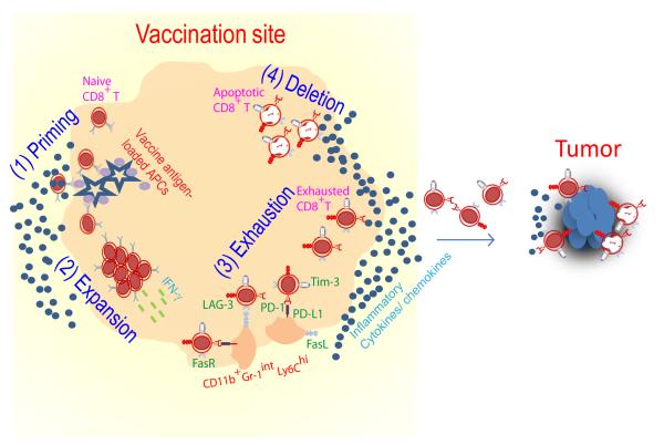
Fig. 1. Fate of CD8+ T cells primed by IFA-based vaccine.
(1) Naive CD8+ T cells travel to antigen-rich and highly inflamed vaccination site and become primed. (2) Effector CD8+ T cells respond to persistent antigen presentation by continued expansion and secretion of inflamma tory cytokines (IFN-γ). (3) Activated CD8+ T cells up regulate Fas and other inhibitory surface markers including PD-1, LAG-3, Tim-3 and CTLA-4 in response to antigen, IFN-γ and other inflammatory cytokines; these conditions also promote accumulation of host cells expressing PD-L1 and FasL. PD-1/PD-L1 engagement leads to T cell exhaustion; Fas/FasL engagement results in T cell apoptosis. (4) As a result, very few primed T cells reach the tumor.

