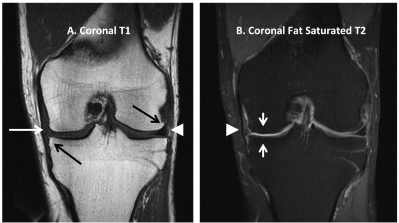
Figure 1.
Representative coronal plane MR images of the knee with examples of pathologic findings and normal morphologic findings. On both images, the left side is medial and the right side is lateral. A. Coronal T1-weighted image with medial tibial plateau osteophyte and lateral femoral condyle osteophyte (black arrows), non-displaced tear of the medial meniscus (white arrow), and normal morphology lateral meniscus (white arrowhead). B. Coronal fat saturated T2-weighted image of the same subject with severe thinning of articular cartilage in the central region of the medial femoral condyle and medial tibial plateau (short white arrows); and normal morphology medial collateral ligament (white arrowhead).

