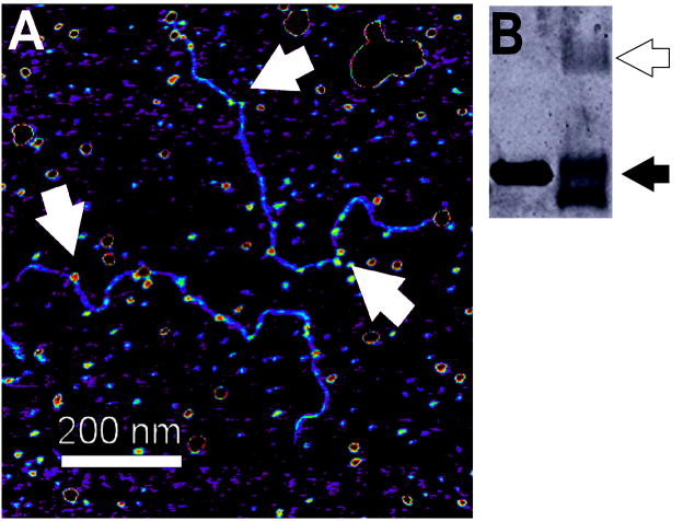
Figure 10. Crosslinked AGT-DNA complexes.
(A) Samples containing linear pUC19 (60 nM) and AGT (12μM) were crosslinked with 0.1% glutaraldehyde (10 min, 37°C) before application to the mica substrate. Arrows indicate AGT clusters on DNA. (B) SDS-PAGE analysis showed that >50% of AGT molecules were crosslinked to a neighbor by this treatment (black arrow monomer, white arrow dimer).

