
Figure 1.
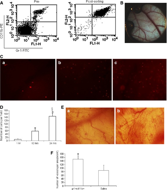
Gr-1+CD11b+ myeloid cells homed to the site of injury after intravenous administration and increased neoangiogenesis at the site of injury in non-diabetic mice. (A) flow cytometry analysis of Gr-1+CD11b+ myeloid cells pre- and post-sorting, which showed the purity of the Gr-1+CD11b+ myeloid cells is ∽95%; (B) establishment of the dorsal window chamber in mice; (C) Gr-1+CD11b+ myeloid cells homed to the site of window chamber injury after intravenous tail vein injection. (a) Gr-1+CD11b+ myeloid cells homed to the site of window chamber 1 hr after injection; (b) Gr-1+CD11b+ myeloid cells homed to the site of window chamber 12 hrs after tail vein injection; (c) Gr-1+CD11b+ myeloid cells homed to the site of window chamber 24 hrs after tail vein injection. Magnification ×40 for each image. (D) With the increasing of time, the number of Gr-1+CD11b+ myeloid cells homing to the site of injury significantly increased. (E) Neoangiogenesis at the window chamber after intravenous injection of Gr-1+CD11b+ myeloid cells. (a) saline treatment; (b) Gr-1+CD11b+ myeloid cells treatment. Magnification ×40 for each image. (F) Gr-1+CD11b+ myeloid cells significantly increased neoangiogenesis at the site of injury, *P < 0.01 versus 1 hr; §P < 0.01 versus 1 and 12 hrs, n = 5 in each group.