
Figure 3.
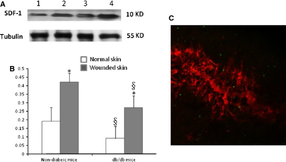
SDF-1 expression at the diabetic wound site was down-regulated compared with non-diabetic mice; however, SDF-1 expression was increased compared with its basal level. (A) Western blot detection of SDF-1 expression at the site of injury. Lane 1, normal skin tissue of diabetic mice; lane 2, skin tissue of diabetic mice at the site of the window chamber; lane 3, normal skin tissue of non-diabetic mice mice; lane 4, skin tissue of non-diabetic mice at the site of the window chamber. The experiments were repeated three times. (B) Densitomery of SDF-1 expression at the injury site 24 hrs after the window chamber model was established; *P < 0.01 versus normal skin; §P < 0.05 versus non-diabetic mice, n = 5 in each group. (C) Gr-1+CD11b+ myeloid cells home to the site of injury after intravenous administration. Red colour indicates the microvessels injected with dextran tetramethylrhodamine imaged with confocal microscope at the site of injury, green colour indicates the Gr-1+CD11b+ myeloid cells with GFP homing to the site of injury; magnification ×40.