
Figure 5.
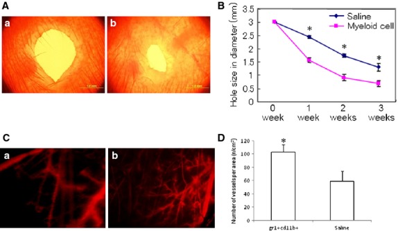
Intravenous injection of Gr-1+CD11b+ myeloid cells enhanced wound healing in diabetic mice. (A) Representative wound size of ear 3 weeks after ear-punch injury. (a) wounded ear from mice intravenously injected with saline; (b) wounded ear from mice intravenously injected with Gr-1+CD11b+ myeloid cells; scale bar = 1 mm. (B) Time course of ear wound healing. Gr-1+CD11b+ myeloid cells significantly enhanced wound healing compared with saline control, *P < 0.01, n = 5 in each group. (C) Representative imaging of neoangiogenesis in the ear 3 weeks after ear-punch injury (red colour indicated the blood vessels filled with dextran tetramethylrhodamine). (a) wounded ear from mice intravenously injected with saline; (b) wounded ear from mice intravenously injected with Gr-1+CD11b+ myeloid cells. Magnification ×40 for each image. (D) Gr-1+CD11b+ myeloid cells significantly increased neoangiogenesis in the wound ear. *P < 0.01 versus saline, n = 5 in each group.