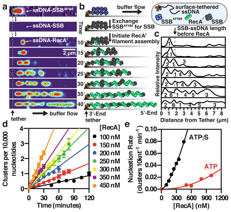
Figure 1. Direct visualization of RecA filament assembly on single molecules of SSB-coated ssDNA reveals that RecA nucleates as a dimer.
(a) RecAf filament assembly with ATPγS on a single molecule of SSB-coated ssDNA tethered within a microfluidic flow chamber was visualized using TIRF microscopy; montage is rendered into a topographic fluorescent intensity map. (b) Schematic and (c) fluorescent intensity profile from panel A. (d) The number of RecA clusters increases linearly with time; slope is the nucleation rate (n = 18–93 clusters for each concentration; (±s.d.)). (e) Nucleation rate increases with [RecA] according to, J=k[RecA]n, where n is 2.2 (±0.6) (ATPγS) and 1.5 (±0.1) (ATP) (error from the linear fits in (d) is smaller than the symbols).

