
Figure 3. Substrate-regulated Hsp70 turnover in vivo and in vitro.
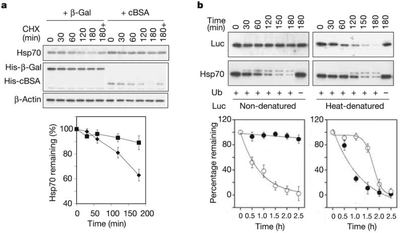
a, HEK-293 cells were co-transfected with plasmids expressing CHIP, Flag-Hsp70 and cBSA (squares) orβ-Gal (diamonds). The turnover of Hsp70 was determined with the use of a cycloheximide (CHX) chase followed by immunoblotting. ‘180 + ’ indicates the presence of 20 μM MG132 during the chase. Values are means ± s.d. b, In vitro degradation assays were performed with Hsp70 (open circles) and luciferase (Luc; filled circles) as substrates. For heat-denatured luciferase, luciferase was heated at 43 °C for 10 min with Hsp70 and Hdj2 (ref. 20). Both the luciferase and Hsp70 levels were determined in the same samples by immunoblotting and quantified by densitometry. Values are means ± s.d.