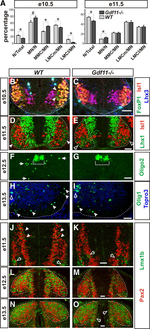
Figure 4.

Slower progression of neurogenesis in the Gdf11−/− embryonic spinal cord. A, Percentage of neurons to total cells (N/Total), motor neurons to neurons (MN/N), and different subtypes of MNs to MNs (MMC/MN, LMCm/MN, and LMCl/MN) in Gdf11−/− and WT littermates at e10.5 and e11.5. *p < 0.05, #p = 0.05. B–G, Images of cross-sectioned ventral spinal cords stained with MN and OLP markers. B, C, White dotted lines delineate the margin of the spinal cord. Arrowheads indicate the presence of late born LMCl MNs (FoxP1+Isl1−Lhx3−) in the WT embryos. Asterisk (*) indicates nonspecific staining. D, E, White dotted lines delineate the ventral horns. Late born LMCl MNs (Lhx1+Isl1−) have migrated through LMCm MNs (Lhx1−Isl1+) to form the LMCl motor column (arrowheads) at the lateral border of the ventral horn in the WT embryo. In the Gdf11−/− spinal cords, the LMCl MNs are still migrating and just starting to reach their final positions (open arrows). F, G, Olig2+ OLPs (white arrows) have started to leave the progenitor domain (white dotted bracket) in WT embryos but not in their Gdf11−/− littermates at e12.5. H, I, Images of right ventral spinal cords, midline is at the left edge of the images and white dotted lines delineate the margin of the spinal cord. Many more Olig1+ OLPs (arrowheads) have migrated into the white matter in the WT compared to the Gdf11−/− embryos. Open arrows indicate the Olig1+ OLPs located in the progenitor domain. J–O, Images of cross-sectioned dorsal spinal cords stained with Pax2 and Lmx1b. J, K, Many Lmx1b+ late-born dILB interneurons (arrowheads) are present in the WT, but few are in Gdf11−/− embryos where Lmx1b+ early born dI5 interneurons (open arrows) are still being generated. L, M, A change in the shape of the dorsal horn (white dotted lines) in WT embryos caused by the influx of Lmx1b+ dILB and Pax2+ dILA interneurons is apparent compared to their Gdf11−/− littermates. N, O, Most of the Lmx1b+ dILB and Pax2+ dILA interneurons have finished their migration into the dorsal horn (white dotted lines) in WT embryos, but many are still being generated and migrating (open arrows) in their Gdf11−/− littermates. Scale bars, 50 μm in all images.
