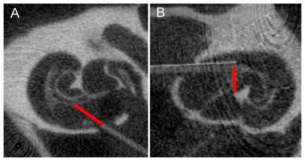
Figure 1.

MicroCT images showing examples of the two different types of fibers, a flat polished fiber (A) and an angle polished fiber (B). The fibers were inserted into scala tympani of a guinea pig. The orientation of the flat polished fiber was towards the modiolus. The angle polished fiber was inserted along scala tympani. The red bar indicates the beam path.
