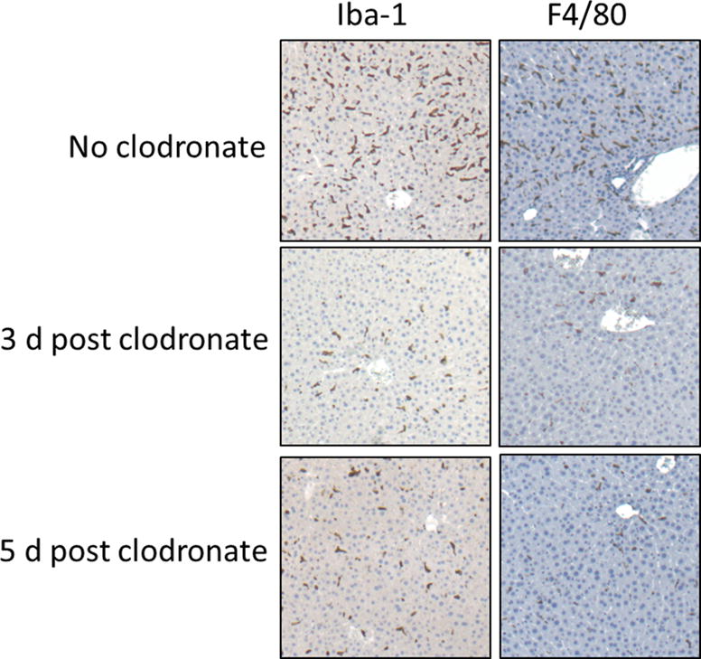
Figure 1.

Immunohistochemical staining of liver sections from huIL-6 mice either not treated or treated with clodronate liposomes 3 or 5 d previously. Staining is shown with mAb Iba1 or mAb F4/80 antibodies both of which are markers for phagocytic cells. Original magnification 50×
