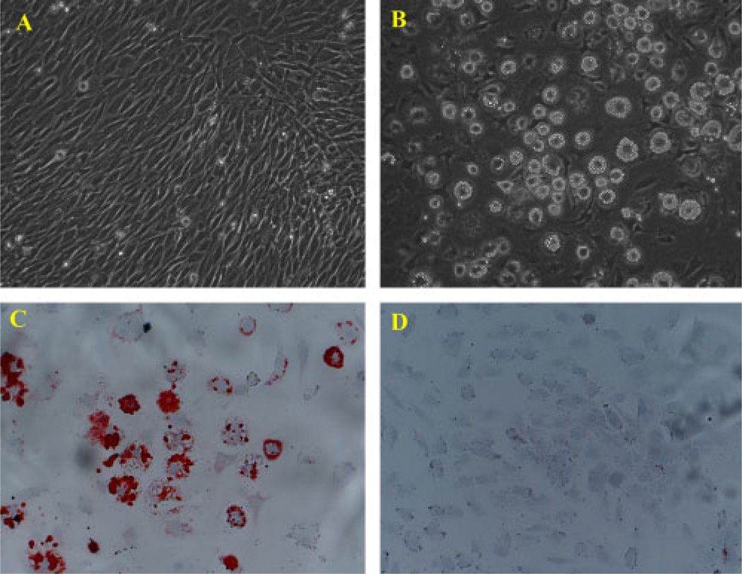
Figure 1.
Morphological change and oil red O staining during adipogenic differentiation of MSC D1 cells. (A) D1 cells in basal culture medium. (B) Morphological change and accumulation of lipid vesicles after D1 cells were treated with adipogenic medium. (C) Cells stained with oil red O for intracellular lipid vesicles after D1 cells were treated with adipogenic medium. (D) oil red O-positive cells were not found in D1 cells in the basal culture medium without inducing agents. Cell nuclei were counterstained with haematoxylin and viewed at × 100 magnification

