
Figure 3. Identification of an intramolecular crosslink in the p22phox subunit of Cyt b.
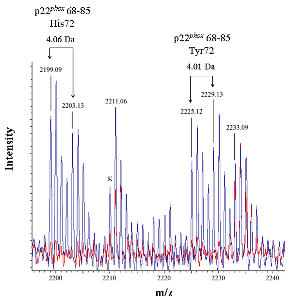
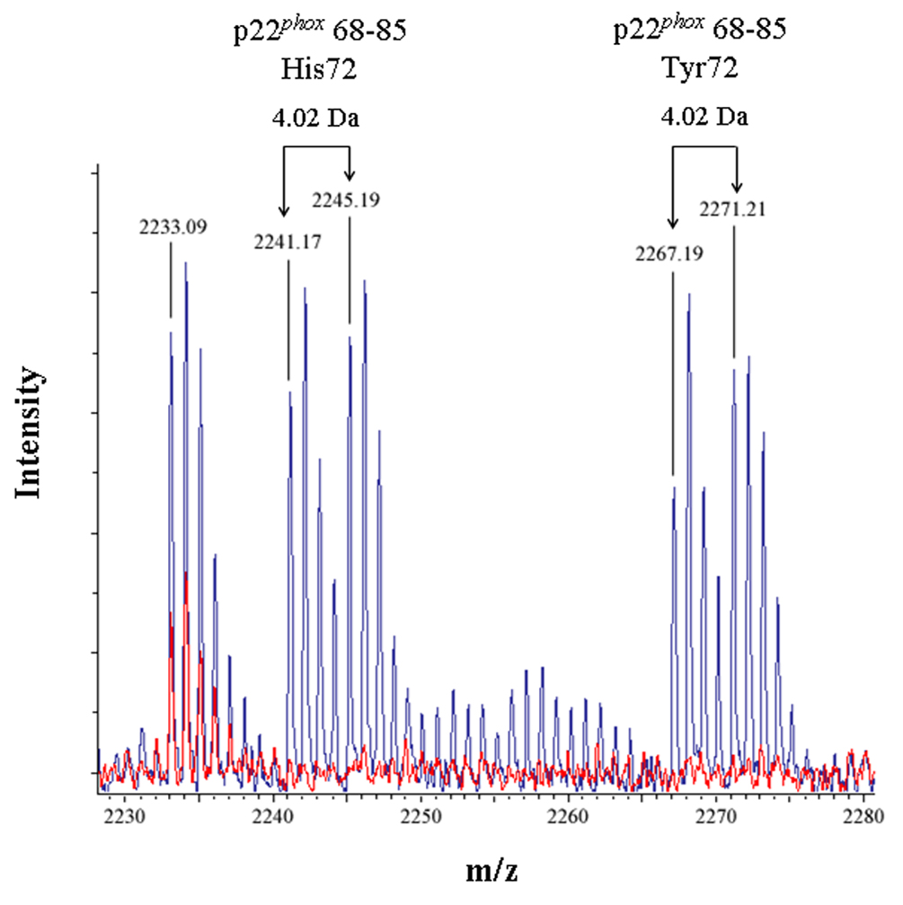
(3A) Overlaid MALDI spectra for Cyt b tryptic digests following incubation with 5% DMSO for 50 min at room temperature (red) and Cyt b tryptic digests following incubation with a 60-fold molar excess of BS2G-d0/d4 for 50 min at room temperature (blue). (3B) Overlaid MALDI spectra for Cyt b tryptic digests following incubation with 5% DMSO for 50 min at room temperature (red) and Cyt b tryptic digests following incubation with a 60-fold molar excess of BS3-d0/d4 for 50 min at room temperature (blue). Masses attributed to gp91phox and p22phox tryptic peptides are labeled numerically in 3A and 3B, and a possible keratin-derived tryptic peptide is indicated (K) in 3B. The initial mass corresponding to the do and d4 peak families in the p22phox crosslink is indicated by arrows. Theoretical masses of p22phox residues 68–85 modified with crosslinking agent are as follows: BS2G-d0 with His72-2199.15 Da; BS2G-d4 with His72-2203.17 Da; BS2G-d0 with Tyr72-2225.15 Da; BS2G-d4 with Tyr72-2229.18 Da; BS3-d0 with His72-2241.20 Da; BS3-d4 with His72-2245.22 Da; BS3-d0 with Tyr72-2267.20 Da; and BS3-d4 with Tyr72-2271.22 Da.