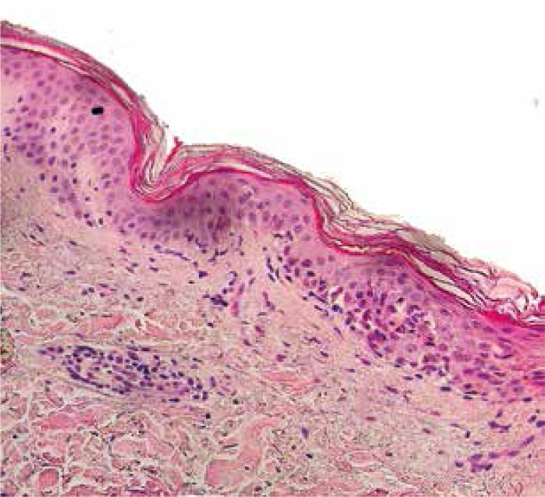
Figure 3.
Histopathological examination of a skin sample taken from the right shoulder area revealed necrotic keratinocytes, vacuolar degeneration in the basal layer, papillary dermal edema and lymphocytic perivascular infiltrates. Keratinocyte necrosis is of rather lower intensity than expected in SJS

