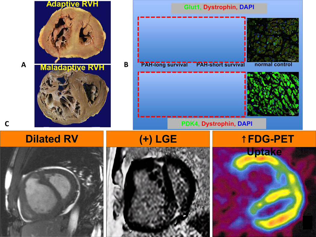
Figure 1.
Increased glycolysis in the right ventricle (RV) in right ventricular hypertrophy (RVH) in PAH patients. (A) The cross sections of RVs from patients with adaptive versus maladaptive RVH. RV chambers are enlarged in both patients however adaptive RVH is concentric with less dilatation and fibrosis. (B) Immunostaining shows up-regulation of Glut1 and PDK4 expression in RV myocytes and is less profound in the PAH patient with adaptive RVH. (C) Imaging modalities showing RV dilatation in MRI, RV fibrosis in MRI and increased FDG uptake in PET scan. The figure is partially adapted from references22, 36, 45with permission.

