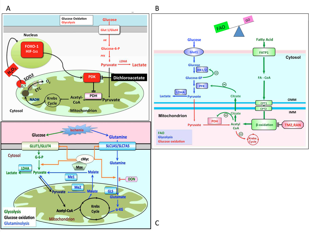
Figure 4.
A: Mechanism of impaired glucose oxidation and enhanced glycolysis in RVH. In RVH, activation of various transcription factors, including FOXO1, cMyc and HIF-1α upregulates expression of many glycolytic gene. A common finding in RVH is increased PDK expression, which inhibits PDH and reduces mitochondrial respiration. PDK activation also occurs in the lung in PAH, although the transcriptional regulation and isoform specificity may differ than that seen in the RV. Dichloroacetate inhibits PDK and thereby promotes glucose oxidation and inhibits glycolysis. ETC = electron transport chain, HK = hexokinase, H2O2 = hydrogen peroxide, LDHA = lactate dehydrogrenase A, PFK = phosphofructokinase. Adapted from 50
B: The Randle cycle in RVH The inhibition of β-FAO by trimetazidine and ranolazine increases PDH activity and improves GO. This reciprocal relationship between GO and FAO is referred to as the Randle cycle. Adapted with permission from 75.
C: Proposed mechanism of glutaminolysis in RVH. RV ischemia and capillary rarefaction activate cMyc and Max, which increases glutamine uptake and production of α-ketoglutarate (α-KG). α-KG enters Krebs’ cycle leading to production of malate. Krebs’ cycle-derived malate generates cytosolic pyruvate, which is converted by lactate dehydrogenase A (LDHA) to lactate. In conditions of high glutaminolysis GO is inhibited. Reproduced from 26 (Illustration Credit: Ben Smith).

