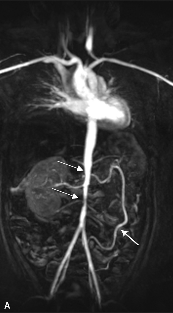
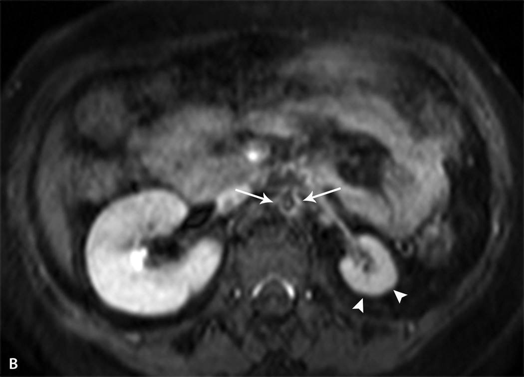
Fig. 5.
Takayasu arteritis. A. Coronal post contrast Maximum Intensity Projection (MIP) MR angiogram in the arterial phase of the chest, abdomen and pelvis in a 13 year-old girl demonstrates narrowing and irregularity of the mid to distal abdominal aorta (thin arrows). There is enlargement of the Arc of Rioland (thick arrow) due to severe stenosis of the celiac and superior mesenteric arteries (not shown). B. Axial post contrast T1 fat-saturated image shows severe narrowing of the abdominal aorta with a thick, enhancing wall (arrows). There is atrophy of the left kidney (arrowheads) due to severe stenosis of the left renal artery.


