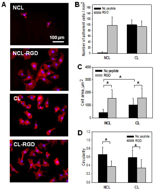
FIGURE 2. Adhesion and spreading of C2C12 myoblasts at early time.
Initial C2C12 cell adhesion and spreading were observed 1 h after plating the cells on NCL, CL, NCL-RGD and CL-RGD films. (A) Actin (red) and nuclei (blue) staining of C2C12 cells to visualize adhesion and spreading on the four types of films (B) Number of adherent cells. (C) Spreading area. (D) Cell circularity quantification. Error bars correspond to SD, *: p < 0.05.

