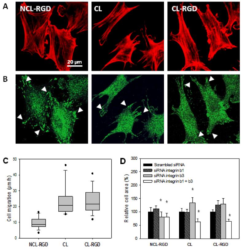
FIGURE 3. Effect of film stiffness and RGD fonctionalization on cytoskeletal organization, focal adhesions and migration.
(A) Staining of actin cytoskeleton (red) after 4 h of culture. (B) Staining of phosphorylated focal adhesion kinase (pFAK Y397, green) after 4 h of culture. (C) Myoblast migration measured over 5 h after seeding. (D) Effect of blocking β1 and/or β3 integrins using siRNA: quantification of the cell area after 4 h of adhesion (* p < 0.05 compared to scrambled siRNA). Focal adhesions/complexes are indicated by white arrowheads.

