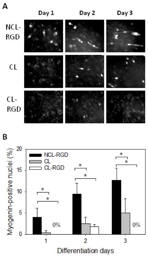
FIGURE 5. Myogenin expression is decreased on stiff films.
After 24 h of proliferation in GM, the medium was changed to DM and cells were let to differentiate for 2 days. (A) Myogenin labeling at day 1, 2 and 3 of differentiation. (B) Quantification of the percentage of myogenin expressing cells. Error bars correspond to SD, *: p < 0.05.

