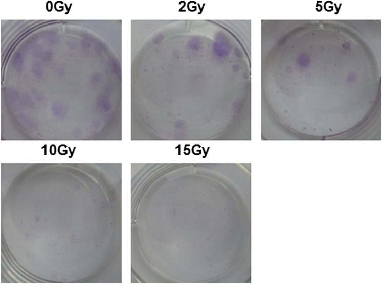
Figure 1.
Characterization of bone marrow mesenchymal stem cells (BMSCs). (a) Histogram of cell surface markers determined by flow cytometry, showing that BMSCs were negative for CD45 and CD34 and positive for CD44 and CD29. (b) BMSCs differentiated into adipocytes and osteoblasts for 14 days and subsequently stained with Oil Red and von Kossa staining, respectively. (c) In vitro irradiation survival curves of human BMSCs (hBMSCs). Cells were grown in vitro and irradiated from 0 to 15 Gy alone. (d) The colony-forming unit fibroblasts (CFU-F) in (c) were counted on the 14th day after ionizing irradiation and presented as CFU-F per 100 cultured cells. (e) Effect of platelet factor (PF4) in irradiation killing of hBMSCs in vitro. Cells were irradiated after 12 h incubation in different doses of PF4. (f) Cumulative administration of PF4. Cells were irradiated after different cumulative administrations of PF4 in vitro. Bars, means ± standard deviation. *p < 0.05; **p < 0.01; #p < 0.001; n = 5.

