
Figure 2.
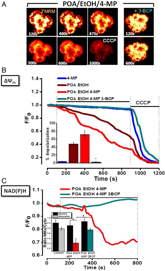
Effects of ethanol (EtOH), palmitoleic acid (POA), 4-methylpyrazole (4-MP) and 3-benzyl-6-chloro-2-pyrone (3-BCP) on mitochondrial membrane potential (ΔψM) in pancreatic acinar cells. (A) Typical fluorescence (glow) images showing progressive loss (six images, left) or maintenance (two images, right) of ΔψM to ethanol/POA/4-MP combination in absence (left), or presence (right) of 3-BCP. Complete mitochondrial depolarisation induced by carbonyl cyanide 3-chlorophenylhydrazone (CCCP;10 μmol/L) is seen at 900 s. (B) Graph showing ethanol/POA-induced loss of ΔψM (n=39), exacerbated by 4-MP (n=44). 4-MP alone was without effect (n=23). 3-BCP abolished effects of ethanol/POA/4-MP (n=36). Summarised data (inset) show mean % depolarisation±SE for each application. (C) Progressive loss of mitochondrial reduced nicotinamide adenine dinucleotide (phosphate) (NAD(P)H) fluorescence induced by ethanol/POA/4-MP (red, n=44) was abolished by 3-BCP (cyan, n=36). Summarised data (inset) show changes of NAD(P)H fluorescence, expressed as mean±SE in mitochondria-specific (mito) versus mitochondria-free (cytosolic; cyto) regions, at baseline (70–100 s) and during stimulation (770–800 s) (* and # p<0.05 compared to control).